Pulmonary Embolism Risk & Symptom Checker
Assess Your Risk Factors
Symptom Checker
Disclaimer: This tool is for educational purposes only and does not replace professional medical advice. If you experience severe symptoms such as sudden shortness of breath, chest pain, or fainting, seek immediate medical attention.
Red Flags: Always consult a healthcare provider if you have any concerns about potential pulmonary embolism, especially after recent surgery, prolonged immobility, or with known risk factors.
Key Takeaways
- A Pulmonary Embolism (PE) occurs when a blood clot blocks a lung artery, often after traveling from the legs.
- Symptoms can be subtle; shortness of breath, chest pain, or sudden faintness should never be ignored.
- Major risk factors include immobility, recent surgery, cancer, and inherited clotting disorders.
- CT pulmonary angiography and D‑dimer testing are the frontline diagnostic tools.
- Anticoagulant therapy-warfarin or direct oral anticoagulants-remains the cornerstone of treatment, with thrombolysis reserved for life‑threatening cases.
What is Pulmonary Embolism?
Pulmonary Embolism is a life‑threatening blockage of a lung artery caused by a blood clot that has traveled from elsewhere in the body, most often from the deep veins of the legs. When the clot lodges in the pulmonary circulation, it reduces oxygen flow and raises pressure on the right side of the heart. The condition can kill within minutes if untreated, which is why it’s dubbed the “silent killer.”
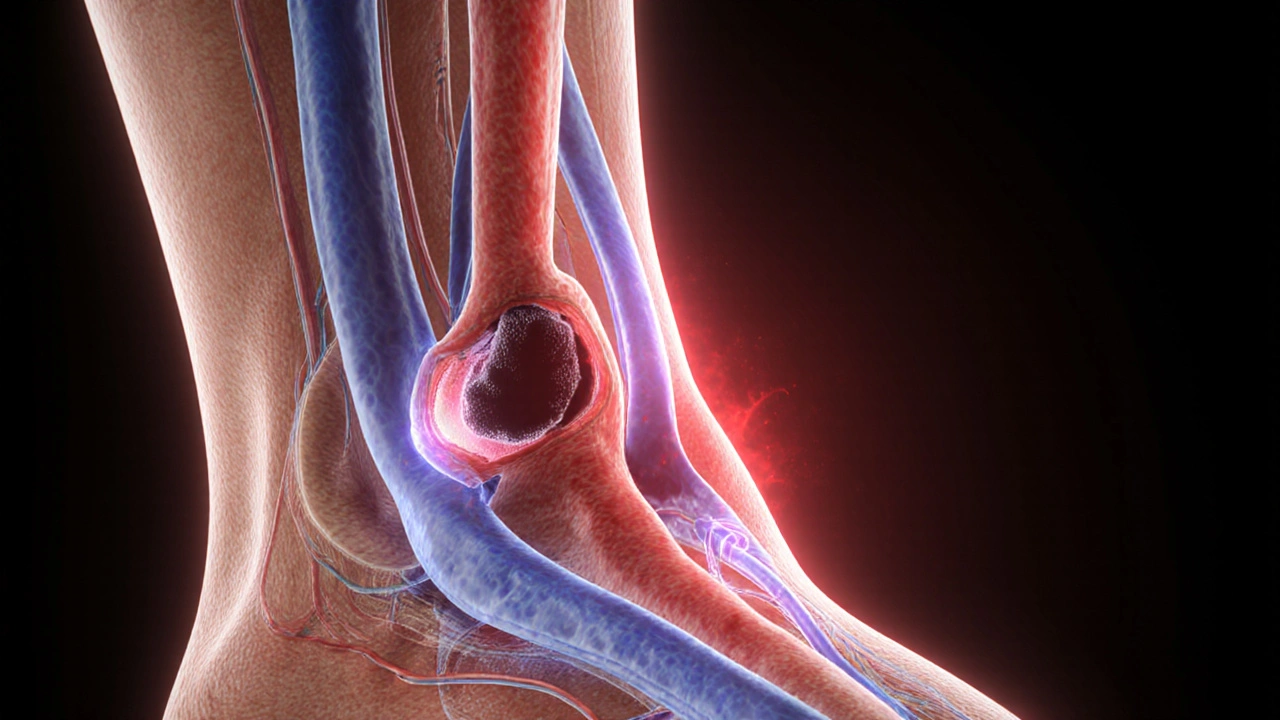
How a Clot Travels: From Leg to Lung
Most PEs start as Deep Vein Thrombosis (DVT), a clot that forms in the deep veins of the calf or thigh. The clot may stay put, but if a piece breaks free it travels through the inferior vena cava, reaches the right atrium, passes into the right ventricle, and is pumped into the pulmonary artery. This journey is called embolization.
Risk factors that encourage clot formation include sluggish blood flow, vessel injury, and a hyper‑coagulable blood state-known as Virchow’s triad.
Who Is at Risk?
Understanding risk helps you stay ahead. Common contributors are:
- Immobility: long flights, prolonged bed rest after surgery, or cast immobilization.
- Recent major surgery, especially orthopedic procedures like hip or knee replacement.
- Cancer and its treatments, which increase clotting factors.
- Hormonal therapy: oral contraceptives or hormone replacement.
- Inherited clotting disorders: Factor V Leiden, prothrombin gene mutation.
- Obesity and smoking, which both elevate blood viscosity.
Even younger, active people aren’t immune-trauma, dehydration, or a family history can tip the balance.
Silent Symptoms and Red Flags
PE often masquerades as a simple cough or anxiety attack. Common clues include:
- Sudden shortness of breath that doesn’t improve with rest. \n
- Sharp, pleuritic chest pain that worsens on deep breathing.
- Rapid heart rate (tachycardia) or feeling of “fluttering” in the chest.
- Light‑headedness, fainting, or a sense of impending doom.
- Swelling or tenderness in one leg-possible DVT sign.
If you notice any of these, especially after a recent surgery or long travel, seek medical attention immediately.
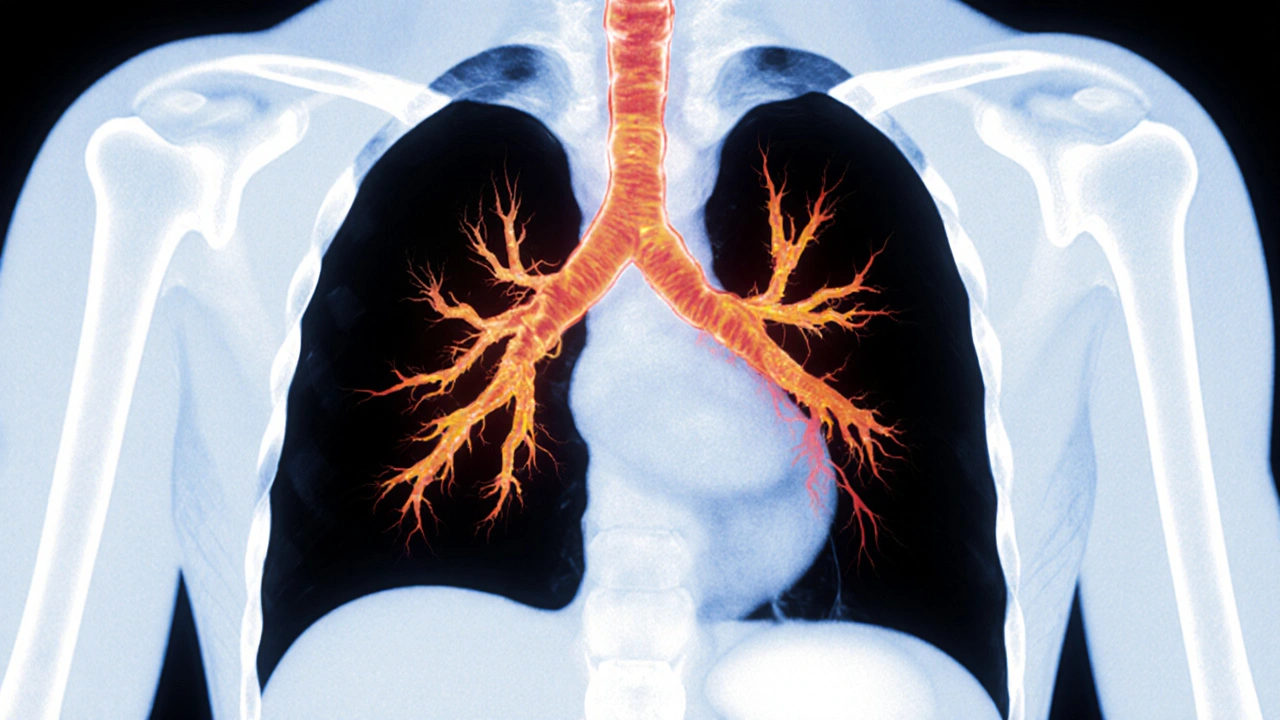
How Is Pulmonary Embolism Diagnosed?
Doctors combine clinical suspicion with objective tests.
- D‑dimer blood test: Elevated levels suggest clot breakdown but are not specific.
- CT pulmonary angiography (CTPA): The gold‑standard imaging. It uses contrast dye to visualize the pulmonary arteries and pinpoint the clot. CT Pulmonary Angiography provides a three‑dimensional map of the clot’s size and location, guiding treatment decisions.
- Ventilation‑perfusion (V/Q) scan: Helps when radiation exposure is a concern.
- Echocardiography: Detects right‑heart strain in massive PEs.
In low‑risk patients, a negative D‑dimer may rule out PE without imaging.
Treatment Options
Once PE is confirmed, therapy moves fast.
- Anticoagulant therapy: Prevents new clots and stops existing ones from growing. Options include Heparin, Warfarin, and newer direct oral anticoagulants (DOACs) like apixaban.
- Thrombolysis: Intravenous clot‑busting drugs (e.g., alteplase) for massive or hemodynamically unstable PE.
- Catheter‑directed therapy: A catheter delivers clot‑dissolving medication or physically removes the clot.
- Surgical embolectomy: Rare, reserved for cases where medication fails and the patient deteriorates.
Typical treatment duration is three to six months, but chronic risk factors may warrant lifelong anticoagulation.
Comparing Anticoagulants
| Attribute | Warfarin | DOACs (e.g., Apixaban) |
|---|---|---|
| Mechanism | Vitamin K antagonist | Factor Xa inhibition |
| Typical dose | 5mg daily (adjusted by INR) | 5mg twice daily (fixed) |
| Monitoring | Regular INR checks (target 2.0‑3.0) | No routine lab monitoring required |
| Dietary restrictions | Avoid high vitamin K foods (leafy greens) | None |
| Renal considerations | Safe in most renal impairment | Dose reduction needed if CrCl <30mL/min |
| Reversal agents | Vitamin K, prothrombin complex concentrate | Andexanet alfa (specific), PCC (off‑label) |
For most patients without severe kidney disease, DOACs offer convenience-fixed dosing, no INR checks, and fewer food interactions. Warfarin remains valuable for patients with mechanical heart valves or severe renal failure.

Prevention Strategies
Stopping a clot before it forms is the safest approach.
- Stay mobile: Walk every hour on long flights or after surgery.
- Compression stockings: Graduated stockings improve leg venous return.
- Hydration: Dehydration thickens blood, especially in hot climates.
- Medication prophylaxis: Low‑dose heparin or DOACs for high‑risk surgical patients.
- Weight management & smoking cessation: Reduce baseline clotting propensity.
Patients with known clotting disorders often work with hematologists to tailor a lifelong prevention plan.
What to Do If You Suspect PE
- Call emergency services (911 in the U.S.) or your local emergency number.
- Tell the dispatcher about recent surgery, long travel, leg swelling, or sudden breathlessness.
- While waiting, sit upright and try to stay calm-avoid lying flat.
- If you have been prescribed anticoagulants before, take the next scheduled dose unless advised otherwise.
- Provide the medical team with a list of current medications, especially blood thinners.
Early medical intervention dramatically improves survival rates, turning a silent killer into a treatable emergency.
Frequently Asked Questions
Can a small pulmonary embolism heal on its own?
Small clots may dissolve gradually, but doctors still prescribe anticoagulants to prevent growth and new clots. Untreated even a tiny PE can cause long‑term lung damage.
Is a D‑dimer test reliable for ruling out PE?
A negative D‑dimer is very helpful in low‑risk patients and can spare you a CT scan. However, a positive result is nonspecific and must be followed by imaging.
What’s the difference between a PE and a heart attack?
A heart attack (myocardial infarction) is caused by a blocked coronary artery, affecting the heart muscle. A PE blocks a lung artery, impairing oxygen exchange. Both can cause chest pain and shortness of breath, but the underlying cause and treatment differ.
Can I travel by plane after a PE?
Most doctors recommend waiting at least two weeks of stable anticoagulation before flying. Use compression stockings, stay hydrated, and move your legs every hour during the flight.
Are there any natural ways to lower clot risk?
Regular exercise, maintaining a healthy weight, and staying well‑hydrated help blood flow. However, natural measures should never replace prescribed anticoagulants for high‑risk patients.
Bottom Line
Pulmonary embolism may strike without warning, but recognizing risk factors, symptoms, and the urgency of treatment can save lives. Keep moving, stay aware of any sudden breathlessness, and don’t hesitate to call for help. With modern imaging and anticoagulants, most patients recover fully and can return to everyday activities.
Wow you think a checklist replaces actually seeing a doc 😒
I guess the tool is nice for a quick glance but don’t rely on it alone. Look up the actual symptoms and risk factors from a real source. If you’re unsure, call your doctor – they’ll take a better look. Also, staying mobile after surgery really helps, trust me.
The pathophysiology of pulmonary embolism involves thrombus formation in the deep veins, embolization to the pulmonary arterial tree, and consequent ventilation‑perfusion mismatch. Early recognition of dyspnea, pleuritic chest pain, and tachycardia is essential. Diagnostic work‑up typically starts with a D‑dimer assay followed by computed tomography pulmonary angiography when indicated 😊.
Another long article that could’ve been a bullet list.
For individuals seeking a comprehensive overview, it is advisable to consult peer‑reviewed medical literature in addition to this summary. Consider discussing anticoagulation options with a hematologist if you possess hereditary clotting disorders. Lifestyle modifications, such as maintaining a healthy weight and avoiding prolonged immobility, further mitigate risk.
Pulmonary embolism is a serious condition that demands your attention. The first step is to recognize the warning signs like sudden shortness of breath. Next you should assess whether you have recent risk factors such as surgery or long travel. If any of those apply you must act quickly and seek medical care. Do not try to self‑diagnose with internet tools alone. A doctor will order the proper tests like a D‑dimer and a CT scan. The results will guide the right treatment plan. Anticoagulant medication is often the cornerstone of therapy. In severe cases doctors may use clot‑busting drugs or even surgery. While on anticoagulants you need regular follow up appointments. Keep an eye on any signs of bleeding and report them immediately. Lifestyle changes such as regular exercise and staying hydrated are beneficial. Wear compression stockings if you have a history of deep vein thrombosis. Educate yourself and your family about when to call emergency services. By staying informed and proactive you can reduce the chances of a life threatening event.
People need to stop ignoring red flags like this – it’s just plain irresponsible to think you can survive a clot on your own.
Honestly, it’s astounding, how many ignore the obvious symptoms, the sudden breathlessness, the crushing chest pain, and then, unsurprisingly, they end up in the ICU, which, frankly, could have been avoided, had they taken even a moment to listen to their bodies.
Look, if you’ve been on a long flight, get up, stretch, walk around a bit it can make a huge diff
Hey there, just a heads‑up that staying active isn’t just a boring suggestion, it’s a downright lifesaver, especially after surgeries or those marathon flights, so set a timer, pop up, do a few leg lifts, maybe even a quick dance move, your circulation will thank you big time and you’ll dodge a nasty clot.
Isn’t it fascinating how a tiny clot can remind us of life’s fragility? 🤔 Staying aware transforms fear into empowerment.
Take a moment each day to check your legs for swelling and keep moving; small habits can prevent big problems.
Indeed, the clinical community has long emphasized the importance of early detection; consequently, patients are encouraged to familiarize themselves with both common and atypical presentations; moreover, adherence to guideline‑directed anticoagulation therapy significantly improves outcomes.
Okay, listen up, if you’re thinking about ignoring those chest pains because you’re “too busy,” you’re basically playing Russian roulette with your lungs, and nobody wants that drama.
From a mechanistic standpoint, the embolic event precipitates a cascade of right‑ventricular strain, hypoxemia, and hemodynamic compromise; therefore, swift initiation of anticoagulation is not merely advisable but imperative to mitigate mortality.
Sure, let’s all just ignore the shortness of breath and keep scrolling, because who needs a life, right? 😂
It’s important, though, to remember that everyone’s risk profile is unique, so personalize your approach and don’t hesitate to reach out to a healthcare professional.